Category: Undergraduate Pathology

Answers to Questions Posted after Year 5 UGI Pathology Lecture

what are the key distinguishing factors that determine if a histological sample is dysplastic or cancer?
Invasion through the baseman membrane into the underlying connective tissue ( in the stomach that is into the lamina propria).

what causes squamous cell carcinoma? (ie you referenced it occurs more commonly in developing countries).
Smoking and/ or alcohol are important. HPV infection also has a role.

I think I missed it but what is CLO-IM?
Columnar Line Oesophagus- Intestinal Metaplasia

Is there such thing as chronic oesophagitis? Or does the inflammation just progress to Barrett’s over time?
These is, for example, with chronic gastro-oesophageal reflux. This will increase the risk of metaplasia

Are there any practical tests for determining cag-A status of H. pylori infections and are these tests used clinically at all?
The tests exist but are not used in routine clinical practice

How common is shock due to ulcer haemorrhage?
It depends how sever the haemorrhage is. Most bleeding from ulcers do not produce shock.

is there a genetic component? i.e. Japanese man migrating to England? (if that makes sense)
There is, and you give a good example, but environmental factors are also important. For example, even when people move they may take their diet with them.

why is there a high incidence in specifically in japan?
It is not only Japan that has a very high incidence: South Korea and Mongolia have even higher incidences. There are environmental as well as genetic factors but it is known that chronic gastritis with intesitnal metaplasia is commoner and more severe in these areas.

Can intestinal gastric cancer progress to diffuse?
I don’t think so but I know nothing published about this. Mixed intestinal and diffuse cancers are not uncommon.

are giardia and Whipple’s disease also associated with immunosuppression?
They are (as are almost all infections).

Neuro-endocrine Tumours ( in less than 500 words)

These tumours arise throughout the body but the gastro-intestinal tract (especially the appendix and rectum), followed by the lung (bronchus), are the commonest sites.
The frequency in these sites reflects the relatively large number of neuro-endocrine cells normally found there. Similarly, in the pancreas, which is another common site for neuro-endocrine tumours (NETs), are commoner in the tail than the head because the Islets of Langerhans are present in a higher density in the tail.

NETs were previously called carcinoid tumours but now the term neuro-endocrine tumours is now preferred except in the lung where the original term is still used.

NETs are graded (1-3) according to their rate of proliferation of the tumour cells which can be assessed either by counting mitoses or using an immunohistochemical marker (Ki-67). The higher the rate, the higher the grade and the greater the risk of metastatic disease although there is no clear cut-off level. The tumours in the appendix and rectum almost always never metastasize. Small intestinal NETs are especially likely to do so. Challengingly the secondary tumours may be larger than the primary they arise from which can make it difficult to identify the latter.

In the gastro-intestinal tract the tumours are usually submucosal although they may ulcerate. They may invade through the wall and may reach the peritoneal surface. They are often associated with a marked desmoplastic reaction. Multiple tumours may be seen; in the ileum 40% are multiple. Spread is to regional lymph nodes and the liver.

Histologically, NETS, are composed of relatively uniform cells arranged, in cords or nests, and have granular cytoplasm. The granules contain a protein, chromogranin, which can be used as an immunohistochemical or serum marker for these tumours.

NETs may secrete a wide range of hormones. The carcinoid syndrome is associated with the secretion of serotonin usually by git tumours. It is characterized by diarrhoea and bronchospasm and may be associated with fibrosis involving the right-hand side of the heart which can produce distortion of the valves. It is only seen in tumours which have spread to the liver as otherwise the serotonin is broken down in the liver.

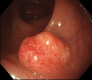
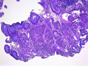
Below is are images of a rectal carcinoid tumour (made available by the excellent @Patholwalker).


Patent Ductus Arteriosus

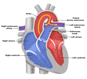
In the fetus, the ductus arteriosus links the pulmonary artery with the aorta. At this time, the pressure in the pulmonary circulation is greater than in the systemic circulation to that blood flows from right to left i.e. from the pulmonary artery into the aorta.

After birth, the ductus close in response to the raised pO2 in the blood and the fibroses up and becomes the ligamentum arteriosus. The ductus may, however, fail to close and this is termed patent ductus arteriosus. This is the 3rd commonest forms of congenital heart disease after ventricular septal defects and atrial septal defects. Like these conditions, it is associated with a left to right shunt (because of the relatively high pressures in the systemic circulation). The magnitude of the shunt will depend on the diameter of the ductus. With time, as a result of the increased blood flow to the lungs, pulmonary hypertension may develop and if the pressure becomes great enough the shunt will reverse becoming right to left (as in the fetus).

This failure to close is common in premature infants when the ductus will usually, in the end, close spontaneously, in the end. Patent ducts arteriosus is commoner in babies with, e.g., Down’s syndrome and congenital rubella. In term infants, it rarely closes on its own. Treatment may be by instilling prostaglandin inhibitors, which cause contraction, or by surgery. Infective endocarditis may develop in untreated cases.

Some Notes on Liver Disease

This was tricky for me to write as the subject is  the closest to my heart! I  really found it hard to leave so much out.


Acute hepatitis:

  1. Viruses
  2. Drugs


Chronic hepatitis (liver disease lasting more than 6 months)

  1. Viruses
  2. Autoimmune
  3. Drugs


Pathological features of chronic hepatitis

Grade = inflammation (can be in 3 places: portal tracts, interface and lobular)

Stage = fibrosis (portal tract expansion > bridging > cirrhosis)


The commonest causes of cirrhosis are:

  1. viral hepatitis
  2. alcoholic liver disease
  3. non-alcoholic fatty liver disease


Viral hepatitis

There are 5 hepatitis viruses (all RNA except for HBV): A-E

A and E: spread by faecal oral route, cause only an acute hepatitis

B, C and D (which can only infect people who have HBV, as well): spread by blood etc., cause the full range of liver disease:

  1. acute hepatitis
  2. chronic hepatitis – scarring begins
  3. cirrhosis – nodules of hepatocytes surrounded by scar tissue


Alcoholic liver disease and non-alcoholic fatty disease (risk factors: obesity, diabetes) produce the same pathological changes:

  1. fatty change
  2. fatty liver hepatitis (alcoholic hepatitis / non-alcoholic steatohepatitis (NASH) respectively): ballooning, neutrophils and scarring
  3. cirrhosis

NB These 3 stages often con-exist


There are (many) other liver diseases which may  cause of cirrhosis:

  1. Autoimmune hepatitis: anti smooth muscle actin autoantibodies, plasma cells, associated with other AI diseases, response to steroids
  2. Drug induced liver injury (DILI): “any kind of liver disease can be caused by a drug’
  3. Haemochromatosis = genetic (AR) increased iron absorption from the gut

deposited in the liver and many other organs (including the pancreas).

  1. Wilson’s disease = genetic (AR) decreased copper excretion (by hepatocytes into the bile duct).
  2. Primary sclerosing cholangitis (PSC): sclerosis (= fibrosis) of the bile ducts leading to their loss. Associated with Ulcerative Colitis
  3. Primary biliary cholangitis (PBC): inflammation (with granulomas) of the bile ducts leading to their loss


Complications of cirrhosis:

  1. Portal hypertension with varices
  2. Liver failure with hepatic encephalopathy,
  3. Liver cell cancer (the same as hepatocellular carcinoma)


Tumours of the liver:

The commonest are secondary tumours (many via the portal vein)


Primary tumours

  1. Benign
  2. Bile duct adenomas
  3. Hepatic adenomas (associated with the contraceptive pill)


  1. Malignant
  2. Liver cell carcinoma

Most commonly associated with cirrhosis.

Spread via the portal vein.

Carry a poor prognosis.


  1. Cholangiocarcinoma (an adenocarcinoma)

Divided into intrahepatic and extra hepatic (including gall bladder)

May be associated with ulcerative colitis and worm infections

Spread to lymph nodes

Carry a poor prognosis


Some Notes on Cardiac Pathology

Please note that I made these for my own use but thought they may be useful to others!

Cardiac Pathology


Divided into diseases of the:

  1. Coronary arteries
  2. Endocardium (including valves)
  3. Myocardium (including congenital heart disease)
  4. Pericardium


  1. Coronary arteries


Any vascular disease can involve these (e.g. vasculitis) but atheroma is the important one.

Clinically: angina, unstable angina and myocardial infarction (due to superimposed thrombosis secondary to ulceration or fissuring).


Left coronary artery >

  • anterior descending > anterior septum and wall of left ventricle
  • circumflex branch > lateral wall of left ventricle


Right coronary artery > posterior septum and wall of left ventricle


Distribution of infarction:

  1. Subendocardial infarction due to severe, generalized disease.
  2. Focal due to blockage of a major artery.


 Complications of myocardial infraction:


Minutes:

Arrhythmias: ventricular fibrillation / heart block

Acute cardiac failure / cardiogenic shock


Days:

Thromboses:

  • Mural (over the infract) which may be followed by systemic embolisation
  • Atrial thrombus (due to atrial fibrillation)
  • (DVT which may be followed by pulmonary embolization)


Week:

Rupture (due to softening of muscle):

  • Myocardium (leading to cardiac tamponade and death)
  • Papillary muscle (mitral incompetence)
  • Septum (left to right shunt)


Pericarditis


Weeks:

  • Chronic cardiac failure.
  • Immune pericarditis (Desslers’s syndrome)


Months:

Cardiac aneurysm (due to fibrosis)


At any time:

Another infarct


  1. B) Myocardium


Myocarditis

Causes:

Infectious:

  • Viral e.g. Coxsackie
  • Bacterial e.g. Borrelia (Lyme Disease)


Toxic: e.g. Diphtheria


Immunological e.g. Rheumatic fever


Cardiomyopathy:

Definition: Heart muscle disease not due to ischaemia, hypertension, valvular disease or inflammation


  1. Dilated cardiomyopathy: end stage of the above (which has burnt out), alcohol or pregnancy
  2. Hypertrophic cardiomyopathy: autosomal dominant
  3. Restrictive cardiomyopathy: endomyocardial, fibro-elastosis, amyloid, haemochromtosis


Rheumatic fever

Preceded by streptococcal sore throat.

Type 2 hypersensitivity reaction (antibodies to streptococci cross react with antibodies to myocardium.


Clinical features

  • General: fever etc.
  • Skin: nodules
  • CNS: chorea
  • Heart:
  • Pericarditis
  • Myocarditis (Aschoff bodies- collections of macrophages)
  • Endocarditis including valves – may lead to chronic valve disease (see below)


Congenital Heart Disease


Risk factors: e.g. Down’s syndrome, rubella, thalidomide


  1. Left to right shunts e.g. atrial or ventricular septal defects ( if untreated may reverse
  2. Right to left shunt “ Cyanotic”

e.g. Tetralogy of Fallot:

  • large ventricular septal defect
  • pulmonary stenosis

3)  overriding of the aorta

4)  right ventricular hypertrophy


  1. C) Endocardium


Valve disease


Mitral valve:


Leads to dilation and hypertrophy of the left atrium

In incompetence there is, also, dilatation of the left ventricle


Incompetence:

  • Post -inflammation: rheumatic fever
  • Infective endocarditis
  • Left ventricular failure
  • Myocardial infarction
  • “Floppy mitral valve syndrome”


Stenosis:

  • Post-inflammation: rheumatic fever


Complications:

  • Atrial fibrillation
  • Infective endocarditis


Aortic valve:


Stenosis:

  • Age related calcification
  • Calcification of abnormal valve:

Congenital bicuspid

Post -inflammation: rheumatic fever


Leads to marked cardiac hypertrophy and the risk of sudden death


Incompetence

  • Post -inflammation: rheumatic fever
  • Infective endocarditis
  • Dilatation of valve ring e.g. Marfan’s syndrome


Leads to dilatation and hypertrophy


Infective endocarditis


Vegetations form on the valves


  1. Acute:

Pathogenic organism (e.g. staphylococcus aureus) and normal valve


  1. Subacute:

Less pathogenic organism (e.g. streptococcus viridans, from the mouth, or enterococci, from the gut) and an abnormal valve


Complications:

  • Systemic features: Fever etc.
  • Embolisation of vegetations

Infected infarcts in the brain or kidneys

Splinter haemorrhages


Other causes of valve vegetations

e.g. marantic in patients with cancer


  1. D) Pericardium


Classified according to appearance

  1. Fibrinous e.g. myocardial infarction
  2. Serous e.g. rheumatic fever
  3. Purulent e.g. bacterial infection
  4. Haemorrhagic e.g. traumatic, tumour
  5. Fibrotic +/- calcification (chronic) = constrictive pericarditis g. TB


Pericardial haemorrhage:

  1. Myocardial infarction
  2. Dissecting aortic aneurysm


Undergraduate Pathology: The Pathology of Neoplasia

This is the text of the first year cellular pathology course lecture which I am giving tomorrow.
I hope that it might act as a focus for interaction with the students.

The Pathology of Neoplasia

Tumour: Any kind of massforming lesion. May be neoplastic (see below), hamartomatous (see below) or inflammatory (e.g. nasal polyps).

Neoplasm: The autonomous growth of tissue which have escaped normal constraints on cell proliferation.
Neoplasms may be either benign (remain localised) or malignant (invade locally and/or spread to distant sites).

Cancers are malignant neoplasms.

Hamartomas are localised benign overgrowths of one of more mature cell types e.g. in the lung. They represent architectural but not cytological abnormalities. For example: lung hamartomas are composed of cartilage and bronchial tissue.

Heterotopias are normal tissue being found in parts of the body where they are no normally present. For example: pancreas in the wall of the large intestine.

Important to note that many malignant tumours rarely cause death (especially skin cancers) and that some benign tumours do kill (usually because of their location, e.g. the brain)

Classification of neoplasms

The primary description of a neoplasm is based on the cell origin and the secondary description is whether it is benign or malignant.

For example, tumours of cartilage are either chondromas (if benign) and chondrosarcomas (if malignant.) The “chondro” stem means derived from cartilage the suffix “oma” means a benign tumour and the suffix ”sarcoma” means a malignant (soft tissue) tumour.

Type of epithelium Benign Tumour Malignant Tumour Example(s)
Squamous Squamous epithelioma or papilloma Squamous cell carcinoma Skin, oesophagus, cervix,

Glandular Adenoma Adenocarcinoma Breast, colon, pancreas, thyroid
Transitional Transitional papilloma Transitional cell carcinoma Bladder

Type of connective tissue Benign Tumour Malignant Tumour Example(s)
Smooth muscle Leiomyoma Leiomyosarcoma Uterus, colon
Bone Osteoma Osteosarcoma
(Osteogenic sarcoma) Arm, leg

Haematological neoplasms Benign Tumour Malignant Tumour Example(s)
Lymphocytes Extremely uncommon Lymphoma Lymphoma
Stomach
Bone marrow Extremely
uncommon Leukaemia Acute lymphoblastic leukaemia,
Chronic myeloid leukaemia

Teratomas

These are tumours derived from germ cells and can contain tissue derive from all three for 3 germ cell layers. They may contain mature and / or mature tissue and even cancers.
Malignant tumours with the suffix “oma”:

1. (Malignant) Lymphoma
2. (Malignant) Melanoma
3. Hepatoma (better called liver cell cancer).
4. Teratoma (not all, see above)

What are the differences between benign and malignant tumours?

1. Invasion: This means direct extension into the adjacent connective tissue and /or other structures e.g. blood vessels. This is what distinguishes dysplasia/ carcinoma in situ from cancer (see next lecture).
2. Metastasis: This means spread via blood vessels etc (see below) to other parts of the body.
NB All malignant tumours have the capacity to metastasise although they may be diagnosed before they have done so
3. Differentiation: This means how much do the cells of the tumour resemble the cells of the tissue it is derived from.
Tumour cells tend to have larger nuclei (and hence a higher nuclear-cytoplasmic ratio) and more mitoses than the normal tissue they are derived from. They may have abnormal mitoses (e.g. tripolar) and marked nuclear pleomorphism (variability in nuclear size and shape).
4. Growth pattern: This means how much does the architecture of the tumour resembles the architecture of the tissue it is derived from.
Tumours have less well defined architecture than the tissue they are derived from.

It is important to note that benign tumours may become malignant.

By which routes do tumours spread?

1. Direct extension. This is associated with a stromal response to the tumour. This includes fibroblastic proliferation (“ a desmoplastic response”), vascular proliferation (angiogenesis) and an immune response.
2. Haematogenous (via blood vessels). The blood vessels usually invaded are the venules and capillaries because they have thinner walls. Most sarcomas metastasise first via the blood vessels.
3. Lymphatic (via lymphatics to lymph nodes and beyond) The pattern of spread is dictated by the normal lymphatic drainage of the organ in question. Most epithelial cancers metastasise first via the lymphatics.
4. Transcoelomic (seeding of body cavities). The commonest examples are the pleural cavities (for intrathoracic cancers) and the peritoneal cavities (for intra-abdominal cancers)
5. Perineural (via nerves) This is an underappreciated route of cancer spread.

How do we assess tumour spread?

1. Clinically
2. Radiologically
3. Pathologically

How do we describe tumour spread (stage)?

T= Tumour: the tumour size or extent of local invasion
N= Nodes : number of lymph nodes involved
M = Metastases: presence of distant metastases

This is called the TNM system and the details are different for each kind of cancer

Grade = how differentiated is the tumour (see Differentiation, above)?
Stage = how far as the tumour spread (see TNM above)?

In terms of tumour prognosis, Stage is more important than Grade.