Category: Gastro-intestinal Pathology

Answers to Questions Posted after Year 5 UGI Pathology Lecture

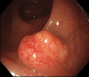
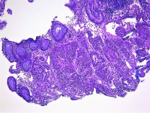
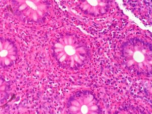
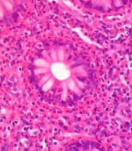
what are the key distinguishing factors that determine if a histological sample is dysplastic or cancer?
Invasion through the baseman membrane into the underlying connective tissue ( in the stomach that is into the lamina propria).

what causes squamous cell carcinoma? (ie you referenced it occurs more commonly in developing countries).
Smoking and/ or alcohol are important. HPV infection also has a role.

I think I missed it but what is CLO-IM?
Columnar Line Oesophagus- Intestinal Metaplasia

Is there such thing as chronic oesophagitis? Or does the inflammation just progress to Barrett’s over time?
These is, for example, with chronic gastro-oesophageal reflux. This will increase the risk of metaplasia

Are there any practical tests for determining cag-A status of H. pylori infections and are these tests used clinically at all?
The tests exist but are not used in routine clinical practice

How common is shock due to ulcer haemorrhage?
It depends how sever the haemorrhage is. Most bleeding from ulcers do not produce shock.

is there a genetic component? i.e. Japanese man migrating to England? (if that makes sense)
There is, and you give a good example, but environmental factors are also important. For example, even when people move they may take their diet with them.

why is there a high incidence in specifically in japan?
It is not only Japan that has a very high incidence: South Korea and Mongolia have even higher incidences. There are environmental as well as genetic factors but it is known that chronic gastritis with intesitnal metaplasia is commoner and more severe in these areas.

Can intestinal gastric cancer progress to diffuse?
I don’t think so but I know nothing published about this. Mixed intestinal and diffuse cancers are not uncommon.

are giardia and Whipple’s disease also associated with immunosuppression?
They are (as are almost all infections).

Neuro-endocrine Tumours ( in less than 500 words)

These tumours arise throughout the body but the gastro-intestinal tract (especially the appendix and rectum), followed by the lung (bronchus), are the commonest sites.
The frequency in these sites reflects the relatively large number of neuro-endocrine cells normally found there. Similarly, in the pancreas, which is another common site for neuro-endocrine tumours (NETs), are commoner in the tail than the head because the Islets of Langerhans are present in a higher density in the tail.

NETs were previously called carcinoid tumours but now the term neuro-endocrine tumours is now preferred except in the lung where the original term is still used.

NETs are graded (1-3) according to their rate of proliferation of the tumour cells which can be assessed either by counting mitoses or using an immunohistochemical marker (Ki-67). The higher the rate, the higher the grade and the greater the risk of metastatic disease although there is no clear cut-off level. The tumours in the appendix and rectum almost always never metastasize. Small intestinal NETs are especially likely to do so. Challengingly the secondary tumours may be larger than the primary they arise from which can make it difficult to identify the latter.

In the gastro-intestinal tract the tumours are usually submucosal although they may ulcerate. They may invade through the wall and may reach the peritoneal surface. They are often associated with a marked desmoplastic reaction. Multiple tumours may be seen; in the ileum 40% are multiple. Spread is to regional lymph nodes and the liver.

Histologically, NETS, are composed of relatively uniform cells arranged, in cords or nests, and have granular cytoplasm. The granules contain a protein, chromogranin, which can be used as an immunohistochemical or serum marker for these tumours.

NETs may secrete a wide range of hormones. The carcinoid syndrome is associated with the secretion of serotonin usually by git tumours. It is characterized by diarrhoea and bronchospasm and may be associated with fibrosis involving the right-hand side of the heart which can produce distortion of the valves. It is only seen in tumours which have spread to the liver as otherwise the serotonin is broken down in the liver.

Below is are images of a rectal carcinoid tumour (made available by the excellent @Patholwalker).


Eosinophils

This post was stimulated by a case of eosinophilic colitis I reviewed at an MDT this morning. The images are below.

They show sheets of eosinophils in the lamina propria and infiltrating crypts. They are easily recognised by their bilobed nuclei and prominent red granules.


This was an opportunity to review eosinophils.

Eosinophils are conspicuous in inflammatory reactions triggered by IgE, such as asthma, and by parasites and are increased by TH-2 activation.  IL-5 and GM-CSF increase the production of eosinophils by the bone marrow. They are associated are recruited into the tissue by eotaxins which are CC chemokines.

Eosinophils have 2 types of effector function:

  1. they release toxic granule proteins  (e.g. major basic protein which is toxic to parasites) and free radicals.
  2. they synthesise prostaglandins, leukotrienes and cytokines.

In the context of this case, likely causes include gut parasites, such as schistosomiasis, and allergic reactions to drugs.

If you want to read more try the excellent British Society of Immunology Website: https://www.immunology.org/public-information/bitesized-immunology/cells/eosinophils