“The Snail is a deadly and cunning beast…”
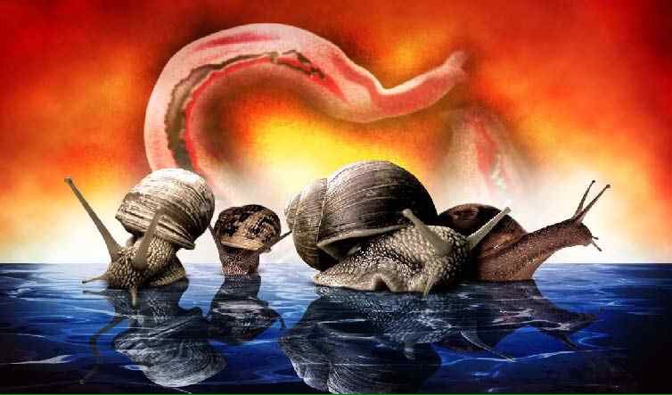
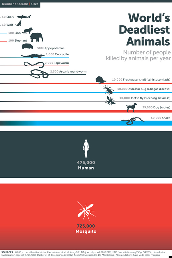
The freshwater snail has been voted within the top 10 most devastating animals to man. Vectors of Schistosomiasis, a condition which currently affects over 260 million people.
Week 2
This week had two main aims:
- Through a “stain and count”procedure determine the most effective wax trap for attracting schistosomes
- Determine what the most effective protein-dye is for dying elastin tissue
Deceptively simple from the outset, these aims proved to be far more difficult to achieve in practice.
The start of our week began in the warm and hospitable environment of the Containment Level 2 Bio-Medical Laboratory at the Wolfson centre in the NHM. Here we received our first batch of patently infected Biomphalaria. Our first few days were spent familiarising ourselves with these poor afflicted Molluscs. Protocol must be respected when dealing with such infectious material; safety naturally becomes a priority and the vitality of the snails must be considered. We moved the snails from dark to light environments to promote heavy shedding – but only permitted shedding for a few hours – after which the snails were returned to dark environments. The snails were fed several times a week and kept in fresh mineral water.
We took samples of this mineral water after a few hours of shedding and used these samples as a source of free cercariae in solution. Cercariae can live for up to 24 hours without finding a host, after this time they will starve and sink. From these samples we were able to practice counting and viewing the cercariae under a field microscope, we also calculated a dilution required to achieve under 100 cercariae in a 1ml sample. From our infected batch of 36 patent Snails we decided a dilution of 1 in 5 (haz:min) was adequate.
Optimising the wax trap
We then assembled our range of wax traps to be submerged in infected water. Based on previous studies we decided that 20 minutes should be enough to promote chemotaxis and burrowing by the cercariae. After this time we planned to retrieve the wax and prepare it with a Lugol (Iodide) protein stain for use under the microscope. We would then simply count the number of stained cercariae in the wax.
Unfortunately this did not work out as expected. This was largely due to two factors:
- Although chemotaxis was well evident within the cercariae toward the wax, the cercariae were not as eager to burrow. We did not witness any direct evidence of penetration into the wax when observing the process live through the microscope.
- Wax is white and opaque, it is very hard to see if cercariae did burrow into wax as they are colourless. The Lugol stain did not diffuse through the wax well.
Slightly deflated, we discussed what could be done to remedy these issues. We decided that a higher ratio of oil to wax should be used to reduce the solidity of the wax and allow it to be spread more thinly across a microscope slide. Other mediums such as agar could also be tried in place of the wax component of the trap. A longer burrowing time should also be allowed – the components of our trap are a poor mimicry of the bio-chemistry of dermal tissue; perhaps the parasites needed longer to take the bait.
These modification would all be made in week 3.
Dying the tissue
We prepared a range of dyes to use on our elastin samples. These included classic protein staining methods using Hematoxylin and Eosin dyes, Fuchsine a common microbiological stain and Acid-wash (wool) dyes.
Unfortunately we experienced a slight delay in receiving our equipment this week so we were missing the Eosin and Acid-wash dyes. Both Hematoxylin and Fuschine could be located in Imperial’s teaching labs however so they were used to prepare our elastin samples. These samples once dyed were subjected to repeated washes, this was to remove any stain left on the surface of the skin samples. After a series of 5 washes the eluted wash water ran clear for both tissue samples.The samples were both still stained heavily red – an indication that the stain had set and dried properly.
The next thing to do was to ensure that these stained tissue samples could be stored over night in water without the stain further dissociating from the tissue. This was achieved with both dyes.
In Week 3 we could then add these stained samples to infected water and see if the activity of elastase promoted the run-off of stain into the surrounding solution. We would also be able to compare the Fuschine and Hematoxylin stains with the Acid-wash and Eosin dyes to see which is best comparing the integrity of stain, response to elastase, and cost.